Stanford researchers accidentally discover a whole new role for the cerebellum
The cerebellum, that weird wrinkly bit of brainstuff at the back of the skull, is sort of the ugly stepsister of the brain. While the cerebrum gets the glory, because it’s the bit we think with, the homely cerebellum toils away unappreciated for its role in motor coordination. But all that is about to change. Scientists from Stanford have stumbled onto a new and different role for the cerebellum — and they found it while they were looking for something else entirely.
“Given what a large fraction of neurons reside in the cerebellum, there’s been relatively little progress made in integrating the cerebellum into the bigger picture of how the brain is solving tasks, and a large part of that disconnect has been this assumption that the cerebellum can only be involved in motor tasks,” said Mark Wagner, who led the research.
Originally, the research team was looking into motor control in mice, to better understand how the cerebellum does what it does. Their target for observation was granule cells, which you can see lit up here in green. Granule cells in the cerebellum account for some 80 percent of the neurons in the brain, numerically, but only 10% of the brain’s volume.
In order to watch how a mouse cerebellum does motor coordination, you need the mouse to move. In this case, the researchers used sugar-water as an enticement to the mice, and watched as the mice pressed a tiny lever to get the sweet reward. Some granule cells lit up as expected, when a mouse was planning and executing arm movements. But other granule cells lit up while the mice were waiting (impatiently, one imagines, oh the tiny mouse frowns) for the sugar water. And others lit up when the researchers took the sugar-water away.
“It was actually a side observation, that, wow, they actually respond to reward,” said lead author Liqun Luo.
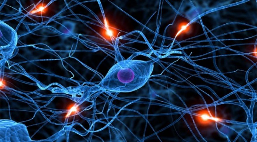
This development was made possible by fluorescence microscopy: a medical imaging technique that uses a dark field so it can pick out things that light up among things that don’t. Fluorescence microscope images are easy to spot, because they’re usually swathes of color on a black background. The things that light up are molecules, each one emitting a pinprick of light as it goes through particular chemical reactions. With calcium imaging, the pinpricks of light come from molecules that fluoresce as they trade calcium ions. Often, they light up in green.
The green is because of biology’s new bestie, GFP: green fluorescent protein. Several species that light up in the dark use GFP to do it, and it turns out the GFP gene is safe and easy to splice into other species’ DNA. As a handy bonus ability, we can splice GFP genes into another creature’s DNA and then watch as the molecule lights up when it’s being translated into RNA or folded into a protein. It sounds tiny, but it lets us watch genes being expressed in real time. Important.
Depending on where in the genome they splice in the GFP gene, scientists can get different bits of a creature to light up under different circumstances. Tools like afford such fine control over splicing that these Stanford scientists were able to tag particular synapses on particular . Since neurons use calcium ions to communicate, neurons are also compatible with calcium imaging. What’s more, calcium imaging can be used in living creatures. No need to dissect and fix a nerve cell. You can watch it fire in real time, right there in situ in the brain — alive and where it belongs.
This involvement in both motor coordination and reward suggests the cerebellum has something important in common with the basal ganglia, a dopamine-driven region of the midbrain that handles reward assessment and resides upstream of the motor and premotor cortices. The basal ganglia also play a part in motor control; Parkinson’s and Huntington’s are both disorders of the basal ganglia that affect coordinated motion. While this discovery about the cerebellum may not lead to a cure for either of those diseases, it does help us to fill out our .